Case #1
-
History: 53 year old female with severe acute onset headache, neck stiffness
© 2012 Must See Radiology

History: 53 year old female with severe acute onset headache, neck stiffness
© 2012 Must See Radiology

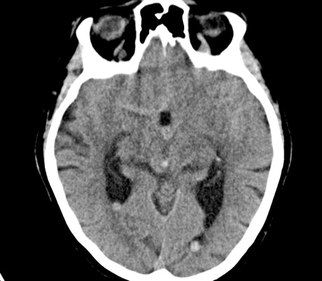
Axial noncontrast CT images through the head at the level of the pons


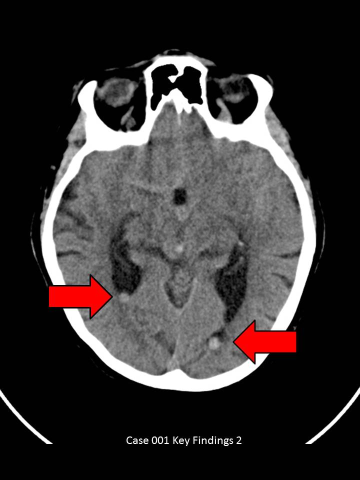
Axial CT images through the brain demonstrate abnormal density in the suprasellar cistern at the level of the pons(yellow circle). Additional dependent collections of high density fluid (hemorrhage) within the bilateral ventricles (red arrows). Also noted is crowding in the foramen magnum with cerebellar tonsil herniation and hemorrhage (not shown above).

Note the CSF density (dark) in the suprasellar cistern. Compare this to the current study
© 2012 Must See Radiology
Subarachnoid Hemorrhage from a Ruptured Basilar Artery Aneurysm
Findings confirmed on cerebral angiogram. The patient underwent coiling of the aneurysm.
Intracranial symmetric hemorrhage from non-traumatic causes can be a difficult diagnosis to make to the untrained eye. Always survey the low-density spaces (cisterns, sinuses, ventricles) for iso/high density blood.
View the prior CT exam from 6 months ago and compare with the current exam. You will notice a higher density (hemorrhage) within the suprasellar cistern on the current study that was not present on the prior.
The suprasellar cistern normally appears as a 5-pointed (pontine level) or 6-pointed (midbrain level) dark star. Also, notice the aneurysmal dilatation of the basilar artery that is present on the prior exam. One of the important findings in this case is the mass effect on adjacent structures. This is seen by cerebral tonsillar herniation and narrowing of the ventricle outflow tract, resulting in mild dilatation of the lateral ventricles.
Additional Information:
Tomandi BF. "CT Angiography of Intracranial Aneurysms: A Focus on Posprocessing." May 2004 Radiographics, 24, 637-655.
© 2012 Must See Radiology
Not available at this time.
Rating not available at this time.
Any feedback regarding this case can be emailed to Tony@mustseeradiology.com
Thank you for trying Must See Radiology!
© 2012 Must See Radiology