Case #18
-
History: 69 year old female with acute onset right sided weakness in the arms and legs.
© 2012 Must See Radiology

History: 69 year old female with acute onset right sided weakness in the arms and legs.
© 2012 Must See Radiology


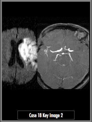
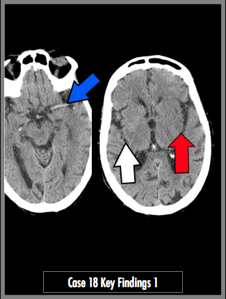
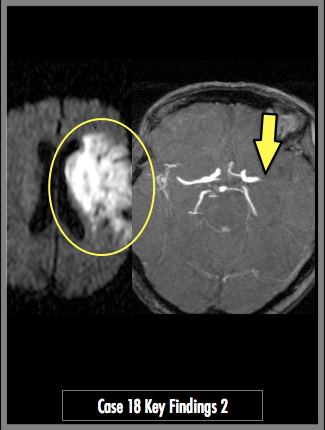
Noncontrast CT of the head (Image 1) and Diffusion Weighted MRI and MRA MIP (Image 2)


CT axial images demonstrate a hyperdense left MCA (blue arrow). Cerebral edema with loss of gray-white matter differentiation in the left insular ribbon (red arrow) compared to the normal right side (white arrow). Large area of diffusion restriction on MRI (yellow circle). Abrupt complete filling defect in the proximal left MCA artery on MRA (yellow arrow)

## ADDL IMAGES ##
© 2012 Must See Radiology
Acute Left Middle Cerebral Artery Infarct
This case is an example of large left MCA territory infarction. These patients present with gross neurologic deficit and it is likely that the ER will be ordering advanced neuro imaging (MRI/MRA or CT perfusion or angiography), even if you miss the subtle findings on CT. The key to the CT is to determine if the infarction is hemorrhagic or not. That is a key piece of information that will affect treatment. This patient did not have a hemorrhagic conversion and had symptoms less than 3 hours, allowing her to be a candidate for thrombolytic therapy.
Additional Information:
Mohr JP "Magnetic resonance versus computed tomographic imaging in acute stroke." Stroke 1995: 26,807-812.
Gonzalez RG "Diffusion-weighted MR imaging: diagnostic accuracy in patients imaged within 6 hours of stroke symptom onset." Radiology 1999: 210,155-162.
© 2012 Must See Radiology
Not available at this time.
Rating not available at this time.
Any feedback regarding this case can be emailed to Tony@mustseeradiology.com
Thank you for trying Must See Radiology!
© 2012 Must See Radiology