Case #25
-
History: Restrained driver hit light pole at 30 MPH. Neck pain. Patient in C-collar.
© 2012 Must See Radiology

History: Restrained driver hit light pole at 30 MPH. Neck pain. Patient in C-collar.
© 2012 Must See Radiology


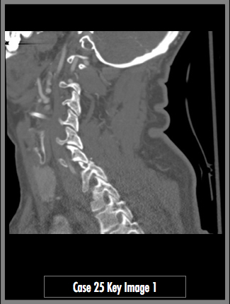
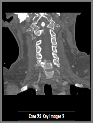
CTA of the neck, sagittal and coronal bone level imaging centered over the cervical spine


Finding 1 demonstrates a unilateral dislocation of the C6 facet (red arrow). There is a fracture of the superior facet articulation of the C7 vertebral body as well (yellow arrow). Note normal facet articulation (white arrow).
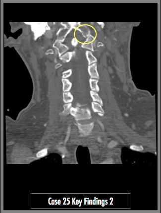
Finding 2 demonstrates a fracture through the left occipital condyle (yellow circle). The fracture is anterior to the left jugular foramen and hypoglossal canal (not pictured).

KEY_FINDING_3_TEXT

KEY_FINDING_4_TEXT

KEY_FINDING_5_TEXT

KEY_FINDING_6_TEXT

KEY_FINDING_7_TEXT
© 2012 Must See Radiology
Unilateral Facet Dislocation / Fracture
& Occipital Condyle Fracture
Method of injury is forced flexion and rotation. Unilateral dislocations are stable, but bilateral dislocations are unstable. These are best demonstrated on the lateral and oblique radiograph / CT views. Anterior dislocation of the affected ody is usually less than half of the AP diameter of the vertebral body. See Primer of Diagnostic Imaging, 5th ed, page 274-275
Occipital Condyle Fractures:
These fractures are best viewed on coronal CT images. They can be subtle and overlooked. If large enough, they can involve the jugular foramen or hypoglossal canal. The jugular foramen contains the inferior petrosal sinus, sigmoid sinus, posterior meningeal artery and cranial nerves #9,10,11. The hypoglossal canal contains cranial nerves #12.
Remember... satisfaction of search is a thing. Don't do it. Learn a search pattern and stick with it, no matter what you find along the way!
Additional Information:
Scher AT "Unilateral Locked Facet in Cervical Spine Injuries." AJR 1977: 129 45-49.
© 2012 Must See Radiology
Not available at this time.
Rating not available at this time.
Any feedback regarding this case can be emailed to Tony@mustseeradiology.com
Thank you for trying Must See Radiology!
© 2012 Must See Radiology