Case #32
-
History: Acute sharp pelvic pain. Dull pain for last few days, but much worse today. Patient is unsure of last menstrual period. B-hCG is positive.
© 2012 Must See Radiology

History: Acute sharp pelvic pain. Dull pain for last few days, but much worse today. Patient is unsure of last menstrual period. B-hCG is positive.
© 2012 Must See Radiology


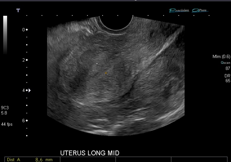
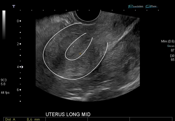
Longitudinal gray scale transvaginal sonographic image of the uterus (left side image). Transverse color Doppler transvaginal sonographic image of the left adnexa (right side image).

Transvaginal image through the uterus (white outline) demonstrates no signs of intrauterine pregnancy. The uterus itself appears normal, however is surrounded in echogenic debris (surrounding entire uterus). Also view the dedicated RUQ and LUQ images in the Case Viewer to see more echogenic debris, representing hemorrhage, in the upper abdomen as well.

Transvaginal images through the left adnexa demonstrate echogenic debris and increased vascularity. No definite gestational sac, yolk sac or embryo identified. Left ovarian follicle present. Don't confuse follicles for ectopic pregnancies in the ovaries.

Transabdominal sonographic color Doppler image through the left adnexa demonstrating echogenic debris (hemorrhage) and increased vascularity.
© 2012 Must See Radiology
Ectopic Pregnancy
Type 3: Ruptured with blood in pelvis
Findings associated with Ectopic Pregnancy:
In the setting of no intra uterine pregancy and/or clinical presentation of ectopic pregnacy:
Types of Ectopic Pregnancy:
Note, these types of ectopic pregnancy, correspond with the above associated US findings
Locations for Ectopic Pregnancy:
Treatment for Ectopic Pregnancy:
Additional Information:
Lin, EP. "Diagnostic Clues to Ectopic Pregnancy." Radiographics 2008: 28, 1661-1671, Last, FM. "Article." Journal Year: ISSUE, PGS.
Bolaji, I. "Sonographic Signs in Ectopic Pregnancy: update." Ultrasound 2012: 20-4, 192-210.
© 2012 Must See Radiology
Not available at this time.
Rating not available at this time.
Any feedback regarding this case can be emailed to Tony@mustseeradiology.com
Thank you for trying Must See Radiology!
© 2012 Must See Radiology