Case #33
-
History: Chest Pain, shortness of breath.
© 2012 Must See Radiology

History: Chest Pain, shortness of breath.
© 2012 Must See Radiology


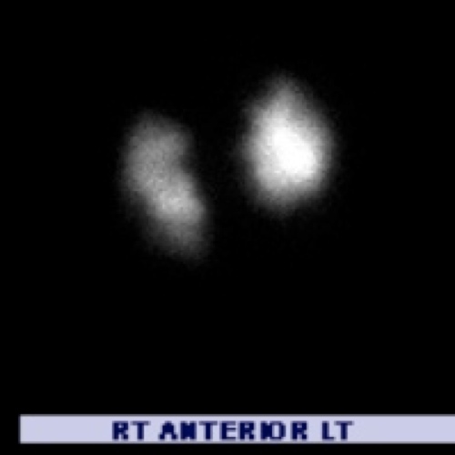
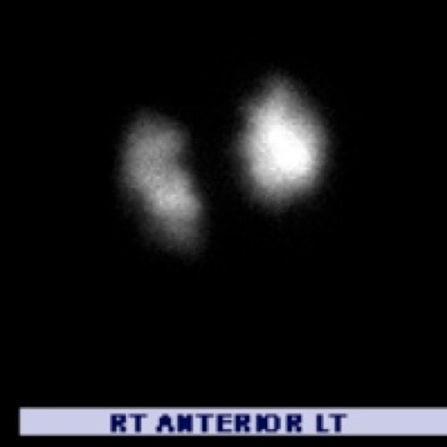
Perfusion (left) and Ventilation (right) in the Anterior projection.


There are multiple large filling defects in the upper and lower lobes of both lungs on the perfusion scan. These are not matched on the ventilation images, and are considered large mismatched defects. There are no radiographic findings to account for the patient's mismatched perfusion defects (see CXR in Case Images). Compare the current study with the prior normal study (available in Case Images)
© 2012 Must See Radiology
High Probability for Pulmonary Embolism
Additional Discussion regarding pulmonary embolism in case #13 and #14
See Case #13 for a more subtle case. See Case #14 for a similar case on CT.Additional Information:
Sostman, HD. "Acute Pulmonary Embolism: Sens and Spec of Ven-Perf Scintigraphy in PIOPED II Study." Radiology Jan 2008, 246:941-946.
Stein, PD. "Contrast enhanced multidetector spiral CT of the chest and lower extremities in the suspected acute pulmonary embolism: results of the PIOPED II." NEJM 2006, 354:2317-2327.
The PIOPED Investigators. "Value of the ventilation/perfusion scan in acute pulmonary embolism: results of the Pulmonary Embolism Diagonsis(PIOPED)". JAMA 1990, 263: 2753-2759.
Wittram C. "How I do it: CT pulmonary angiography." AJR 2007, 188: 1255-1261.
© 2012 Must See Radiology
Not available at this time.
Rating not available at this time.
Any feedback regarding this case can be emailed to Tony@mustseeradiology.com
Thank you for trying Must See Radiology!
© 2012 Must See Radiology